Αναζήτηση αυτού του ιστολογίου

Κυριακή 3 Απριλίου 2022

Development and Validation of Instrument for Operative Competency Assessment in Selective Neck Dissection

paythelady.61 shared this article with you from Inoreader
Annals of Otology, Rhinology &Laryngology, Ahead of Print.
Background:Instruments to assess surgical skills have been validated for several key indicator procedures in otolaryngology. Selective neck dissection is a core procedure for which trainees must integrate knowledge of complex head and neck anatomy with technical surgical skills. An instrument for assessment of surgical performance in selective neck dissection has not been previously developed. The objective of the current study is to develop and validate an instrument for assessing surgical competency for level II-IV selective neck dissection.Design:A Delphi working group comprised of 23 fellowship trained head and neck surgeons from 17 institutions was assembled. The modified Delphi method encompassed a 3-step process, including 2 anonymous voting rounds to successively refine individual items and establish levels of consensus. Thresholds for achieving strong consensus, at >80% agreement, were determined a p riori. The resulting instrument was subsequently validated in a prospective cohort of 17 resident surgeons, spanning postgraduate year 1 to 5 training experience. Participants were asked to perform a level II-IV selective neck dissection on fresh-frozen cadaveric specimens. Performance was scored by 2 independent, blinded observers using the devised instrument and construct validity was assessed.Results:Through the modified Delphi process a final list of 30 items, considered to be the most essential items for achieving the goals of a level II-IV selective neck dissection, was developed. Construct validity was supported by a positive association between instrument scores compared to both resident postgraduate year level and number of head and neck rotations completed.Conclusion:The development and validation of a novel instrument for assessment of surgical competency in level II-IV selective neck dissection, a key indicator case in otolaryngology, is described. This new instrument ma y be used to provide objective feedback on overall and task-specific competency to identify surgical deficiencies and offer granular feedback to enhance surgical training.
View on the web

Readmission Rates Following Major Head and Neck Surgery With Concurrent Tracheostomy

paythelady.61 shared this article with you from Inoreader
Annals of Otology, Rhinology &Laryngology, Ahead of Print.
Objectives:To determine the influence of major head and neck procedures on readmission and complication rates following tracheostomy.Methods:A retrospective cohort study using the 2005 to 2017 National Surgical Quality Improvement Program (NSQIP) database. Current Procedural Terminology codes were used to identify tracheostomy patients and to define the underlying head and neck procedure. Patients under the age of 18 and with unknown pre-operative variables were excluded. Univariate and multivariable analyses were performed.Results:A total of 3240 tracheostomy patients undergoing major head and neck surgery were identified in NSQIP. The 30-day mortality rate was 104 (3.2%) and 258 (9.0%) patients were readmitted. 637 (19.7%) patients had an unplanned return to the operating room. There were 1606 (49.6%) non-tracheostomy specific complications, which included 850 (26.2%) medical and 1142 (35.2%) surgical complica tions. On multivariable analysis, we found that the underlying procedures did not impact the risk of readmission (P > .05 for all). The underlying procedure was also not associated with unplanned return to the operating room except for thyroidectomies, which had a lower risk than free tissue graft reconstruction (OR = 0.53 (95%CI 0.31, 0.88), P = .018).Conclusion:While almost 1 in every 2 patients had a complication following major head and neck surgery that included creation of a tracheostomy, the rate of readmission is comparatively low and is not associated with the underlying procedure. These findings should reassure head and neck surgeons that properly managed tracheostomies do not constitute a disproportionate risk of readmission.
View on the web

The Effect of Topical Tranexamic Acid in Endoscopic Sinus Surgery: A Triple Blind Randomized Clinical Trial

paythelady.61 shared this article with you from Inoreader
Annals of Otology, Rhinology &Laryngology, Ahead of Print.
Objective:Our aim is to evaluate the effect of topical tranexamic acid (TA) on bleeding and surgical quality field in the functional endoscopic sinus surgery (FESS).Methods:A total of 74 patients who underwent FESS due to chronic rhinosinusitis were included. The patients were randomized into 2 groups. TA group (n = 37) received a topical cotton pledget soaked with TA and placebo (PL) group (n = 37) received a pledget soaked with saline solution.Results:A significant effect was noted for the TA group versus the PL group in the grade 1 of the Boezaart scale at 35 minutes (4 for TA group and no case for PL group). This effect was absent for higher grades. We did not notice a significant effect between the 2 groups at 5 minutes. Blood loss was 359 ml in the TA group versus 441 ml in the PL group. No significant change was observed between the 2 groups concerning the blood parameters. No side effects were reported.Conclusion:Despite its safety when administrated locally and its low cost, TA provides limited effect on quality of surgical field after 35 minutes of the start of FESS in the patients with chronic rhinosinusitis. This effect was absent at the start of the intervention and when analyzing the blood loss and hematologic parameters.
View on the web

Cervical Necrotizing Fasciitis and Free Tissue Transfer

paythelady.61 shared this article with you from Inoreader
Annals of Otology, Rhinology &Laryngology, Ahead of Print.
Objectives:Aggressive surgical debridement is required in cervical necrotizing fasciitis, and in severe defects, subsequent free tissue transfer might be necessary. However, there is concern that the inflammatory environment of the infection site may threaten free flap viability, particularly with concerns for thrombosis of feeding vessels and compromised tissue integration. Cases in the head and neck area are rare, so there are limited data regarding outcomes of free tissue transfer in these patients.Methods:A retrospective chart review assessed patients with cervical necrotizing fasciitis treated at an academic tertiary hospital between 2015 and 2021. Twenty-five patients were identified, and eight required free tissue transfer after adequate surgical debridement. Treatment, hospital course, and demographic data were collected on these eight patients.Results:All flaps had full survival at follow up (median fol low up 3 months, range 1-39 months) without concerns for vascular compromise.Conclusion:These data suggest that in patients with large soft tissue defects due to cervical necrotizing fasciitis, free tissue transfer may be a safe treatment modality.
View on the web

Benign paroxysmal positional vertigo

paythelady.61 shared this article with you from Inoreader
Benign paroxysmal positional vertigo (BPPV) is characterized by positional vertigo (brief attacks of rotatory vertigo triggered by head position changes in the direction of gravity) and is the most common peripheral cause of vertigo. There are two types of BPPV pathophysiology: canalolithiasis and cupulolithiasis. In canalolithiasis, otoconial debris is detached from the otolithic membrane and floats freely within the endolymph of the canal. In cupulolithiasis, the otoconial debris released from the otolithic membrane settles on the cupula of the semicircular canal and the specific gravity of the cupula is increased.
View on the web

Πέμπτη 31 Μαρτίου 2022

A Mass in the Infratemporal Fossa

paythelady.61 shared this article with you from Inoreader

jamanetwork.com

A 51-year-old woman presented with headache, jaw pain, paresthesia, trismus, and a history of anxiety, depression, emphysema, hyperlipidemia, migraines, pseudoseizures, and no pertinent surgery nor head trauma. What is your diagnosis?
View on the web

Key Issues in Use of Prophylactic Steroids to Prevent Hypocalcemia and Voice Dysfunction After Thyroidectomy—Reply

paythelady.61 shared this article with you from Inoreader

jamanetwork.com

In Reply We thank Ms Kane and colleagues for their feedback on our article.
View on the web

Disability After Computerized Vestibular Retraining for Stable Unilateral Vestibular Deficit

paythelady.61 shared this article with you from Inoreader

jamanetwork.com

This cohort study evaluates whether vestibular retraining using computerized dynamic posturography is associated with reduced participant-reported disability for patients with an objectively assessed unilateral peripheral vestibular deficit.
View on the web

Vestibular Rehabilitation for Unilateral Peripheral Vestibular Deficits

paythelady.61 shared this article with you from Inoreader

jamanetwork.com

In the 1940s, Cawthorne and Cooksey noted that patients with unilateral vestibular dysfunctions who exercised coped better with their symptoms and made a faster recovery than patients who did not exercise. These original exercise protocols were developed to treat patients with labyrinth injury resulting from head injury or surgery. Vestibular rehabilitation can be defined as a set of exercises designed to facilitate central nervous system plasticity through adaptation or by generating substitute mechanisms among patients with balance disorders, thus improving their overall stability and helping them resume their daily activities. Vestibular rehabilitation has been practiced for almost 70 years and evidence related to its efficacy and effectiveness has increased over the last 15 to 20 years. According to the Cochrane systematic review by McDonnell and Hillier, there is moderate to strong evidence that vestibular rehabilitation (movement, exercise based) is a safe and effective approach for unilateral peripheral vestibular disorders.
View on the web

Evaluation of digital construction, production and intraoral position accuracy of novel 3D CAD/CAM titanium retainers

paythelady.61 shared this article with you from Inoreader

56.jpg

Abstract

Objectives

New opportunities have arisen to manufacture three-dimensional computer-aided design/computer-aided manufacturing (3D CAD/CAM) retainers from titanium blocks by digital cutting technology. These novel technologies need to fulfill requirements regarding digital planning and position accuracy. The aim of the present study was to investigate the digital construction, the CAD/CAM production and the intraoral positioning accuracy of custom-manufactured novel 3D CAD/CAM titanium retainers.

Materials and methods

A total of 37 prime4me® RETAIN3R (Dentaurum, Ispringen, Germany) retainers were inserted to stabilize the upper anterior front teeth. Following insertion, an intraoral scan was used to record the position. The intraoral position was compared to the virtual setup using 3D superimposition software. Measurement points were evaluated in all three dimensions (horizontal, sagittal and vertical planes). Data were analyzed using Kruskal–Wallis test followed by Dunn's multiple comparison test.

Results

A total of 185 measurements were performed. The horizontal plane and the sagittal plane demonstrated a high level of positioning accuracy between the planned and the intraoral position. Statistically significant deviations between the preceding virtual setup and the intraoral situation were observed in the vertical dimension. Within the retainer, the intraoral positioning accuracy decreased for the measurement points in the direction of the distal retainer segment.

Conclusion

Based on the results, the present study shows a high level of congruence between the 3D virtually planning and the final intraoral position of the fabricated novel 3D CAD/CAM titanium retainers.

View on the web

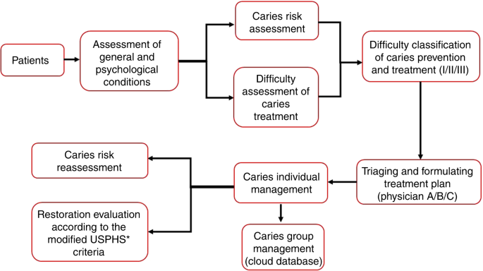
Expert consensus on dental caries management

paythelady.61 shared this article with you from Inoreader

41368_2022_167_Fig1_HTML.png

International Journal of Oral Science, Published online: 31 March 2022; doi:10.1038/s41368-022-00167-3

Expert consensus on dental caries management
View on the web

Τετάρτη 30 Μαρτίου 2022

Comparison of stress distribution between zirconia/alloy endocrown and CAD/CAM multi-piece zirconia post-crown: three-dimensional finite element analysis

paythelady.61 shared this article with you from Inoreader

784.jpg

Abstract

Objectives

This study aimed to evaluate a digital multi-piece zirconia post-crown to restore a mandibular second molar with extensive coronal loss and limited restoration space, and to compare the stress distribution between endocrowns made of zirconia or alloy and CAD/CAM multi-piece zirconia post-crowns.

Material and methods

Four three-dimensional finite element analysis models of a mandibular second molar with extensive coronal loss and limited restoration space were created as follows: (A) intact molar; (B) zirconia endocrown restored molar; (C) multi-piece post-crown restored-molar with tapered nail; (D) multi-piece post-crown restored molar with T-shaped nail. Models C and D were divided into two subgroups according to the material type: C1/D1, zirconia; C2/D2, NiCr alloy. The maximum modified von Mises failure criterion (mvM) stresses were calculated, and the stress distribution was recorded to analyze the effects of the restoration and material types on the biomechanical properties of dentin and prosthesis.

Results

The maximum mvM stress of dentin in model B (33.80 MPa) was lower compared with models C (C1, 37.81 MPa; C2, 36.36 MPa) and D (D1, 36.34 MPa; D2, 34.97 MPa) under vertical load, but the opposite was observed under oblique load. The highest mvM stress was concentrated in the nail region located in the root canal, and the T-shaped nail values were greater than the tapered nail, whereas model D with T-shaped nail showed a lower mvM stress level in dentin compared with Model C with tapered nail.

Conclusions

The digital multi-piece zirconia post-crown is a potential approach to restore mandibular second molars with extensive coronal loss and limited restoration space.

Clinical relevance

The digital multi-piece zirconia post-crown has potential to restore mandibular second molars with extensive coronal loss and limited restoration space using an innovative approach.


View on the web